Tissue engineering in bone regeneration
DOI:
https://doi.org/10.35366/105480Keywords:
Bone regeneration, bone tissue engineering, boneAbstract
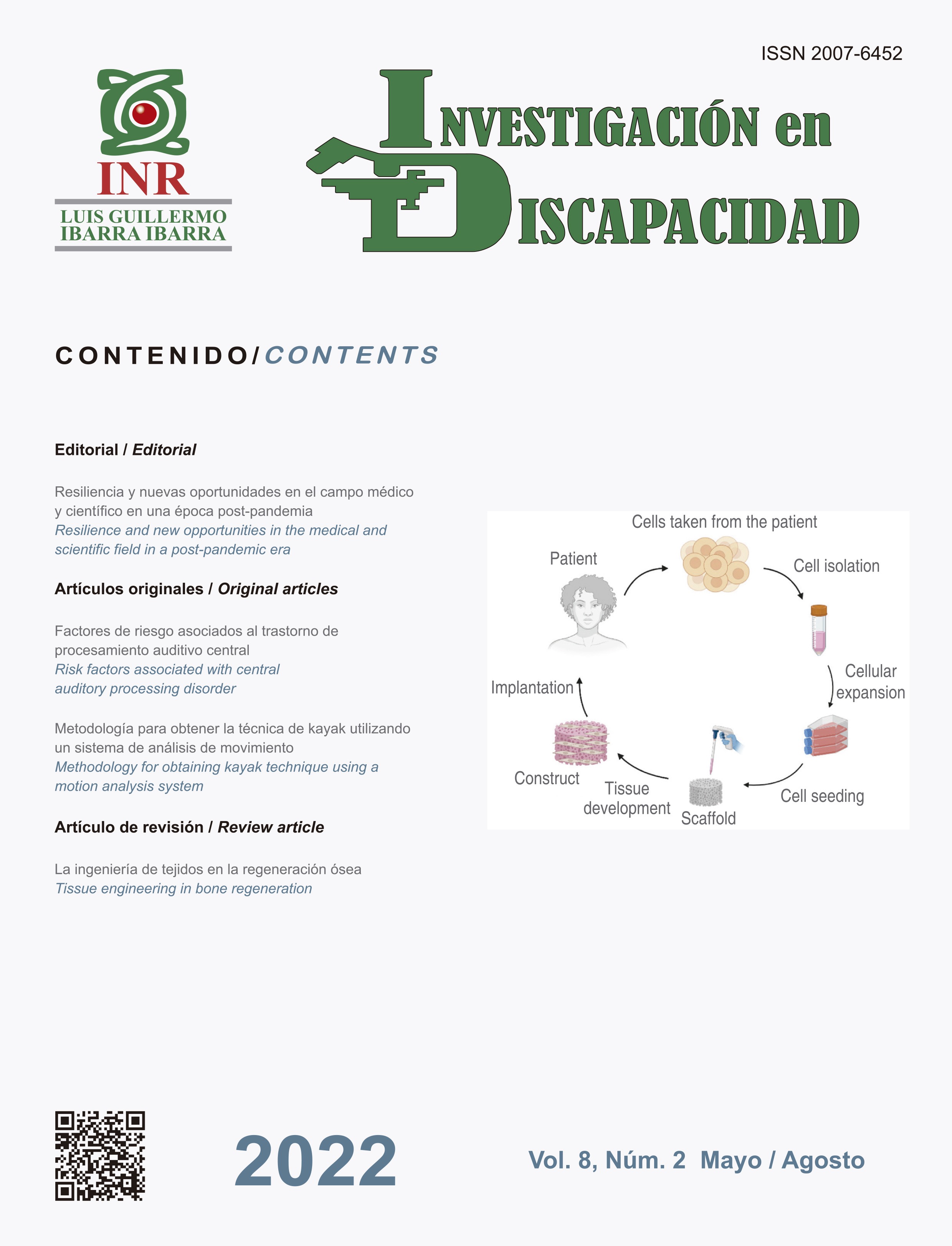
The objective of regenerative medicine is to repair and replace damaged tissues or lost, initiating the process of natural regeneration, and using technologies such as tissue engineering. Bone tissue engineering requires a scaffold, a source of cells and growth factors, alone or in combination, to initiate the process of tissue regeneration. Several studies have developed safe and effective scaffolds for clinical use; some biomaterials used for bone reconstruction include ceramics, demineralized bone matrix, metals, and natural or synthetic biopolymers. The cells are an integral part of the strategy of Tissue Engineering, isolation, expansion efficiency, stability of the osteoblast phenotype, the ability of bone formation in vivo, as well as long-term security are essential requirements that must be met by any osteogenic cell type for successful clinical application in tissue engineering concepts. Growth factors are essential in tissue engineering because they function as signaling molecules that promote or prevent cell adhesion, proliferation, migration, and differentiation. This draft will mention each compound using tissue engineering strategy to repair and regenerate bone lesions and their clinical applications.
Publication Facts
Reviewer profiles N/A
Author statements
Indexed in
- Academic society
- N/A
References
Nicoll SB. Materials for bone graft substitutes and
osseous tissue regeneration. In: Burdick JA, Mauck
RL. Biomaterials for tissue engineering applications.
Philadelphia, Pennsylvania, USA: Editorial Springer Wien
New York; 2011. pp. 343-362.
Peric KZ, Rider P, Alkildani S et al. An introduction to
bone tissue engineering. Int J Artif Organs. 2020; 43 (2):
-86. doi: 10.1177/0391398819876286.
Adel IM, ElMeligy MF, Elkasabgy NA. Conventional and
recent trends of scaffolds fabrication: a superior mode
for tissue engineering. Pharmaceutics. 2022; 14 (2): 306.
doi: 10.3390/pharmaceutics14020306.
Borrelli MR, Hu MS, Longaker MT, Lorenz HP.
Tissue engineering and regenerative medicine in
craniofacial reconstruction and facial aesthetics. J
Craniofac Surg. 2020; 31 (1): 15-27. doi: 10.1097/SCS.
Adamski R, Siuta D. Mechanical, structural, and biological
properties of chitosan/hydroxyapatite/silica composites
for bone tissue engineering. Molecules. 2021; 26 (7):
doi: 10.3390/molecules26071976.
Yu X, Tang X, Gohil SV, Laurencin CT. Biomaterials for
bone regenerative engineering. Adv Healthc Mater. 2015;
(9): 1268-1285. doi: 10.1002/adhm.201400760.
Storti G, Scioli MG, Kim BS, Orlandi A, Cervelli V.
Adipose-derived stem cells in bone tissue engineering:
useful tools with new applications. Stem Cells Int. 2019;
: 3673857. doi: 10.1155/2019/3673857.
Colazo JM, Evans BC, Farinas AF, Al-Kassis S, Duvall
CL, Thayer WP. Applied bioengineering in tissue
reconstruction, replacement, and regeneration. Tissue
Eng Part B Rev. 2019; 25 (4): 259-290. doi: 10.1089/
ten.TEB.2018.0325.
Ho-Shui-Ling A, Bolander J, Rustom LE, Johnson AW,
Luyten FP, Picart C. Bone regeneration strategies:
Engineered scaffolds, bioactive molecules and stem cells
current stage and future perspectives. Biomaterials. 2018;
: 143-162. doi: 10.1016/j.biomaterials.2018.07.017.
Jeyaraman M, Muthu S, Gangadaran P et al. Osteogenic
and chondrogenic potential of periosteum-derived
mesenchymal stromal cells: do they hold the key to the
future? Pharmaceuticals (Basel). 2021; 14 (11): 1133.
doi: 10.3390/ph14111133.
Polini A, Pisignano D, Parodi M, Quarto R, Scaglione
S. Osteoinduction of human mesenchymal stem cells
by bioactive composite scaffolds without supplemental
osteogenic growth factors. PLoS One. 2011; 6 (10):
e26211. doi: 10.1371/journal.pone.0026211.
Thrivikraman G, Athirasala A, Twohig C, Boda SK,
Bertassoni LE. Biomaterials for craniofacial bone
regeneration. Dent Clin North Am. 2017; 61 (4): 835-856.
doi: 10.1016/j.cden.2017.06.003.
Losee JE, Karmacharya J, Gannon FH et al. Reconstruction
of the immature craniofacial skeleton with a carbonated
calcium phosphate bone cement: interaction with
bioresorbable mesh. J Craniofac Surg. 2003; 14 (1): 117-
doi: 10.1097/00001665-200301000-00022.
Yang Y, Kang Y, Sen M, Park S. Bioceramics in tissue
engineering. In: Burdick JA, Mauck RL. Biomaterials
for tissue engineering applications. Philadelphia,
Pennsylvania, USA: Editorial Springer Wien New York;
pp. 343-362.
Zhang W, Yelick PC. Craniofacial Tissue Engineering.
Cold Spring Harb Perspect Med. 2018;8(1):a025775.
doi:10.1101/cshperspect.a025775.
Kang NH, Kim SJ, Song SH, Choi SM, Choi SY,
Kim YJ. Hydroxyapatite synthesis using EDTA. J
Craniofac Surg. 2013; 24 (3): 1042-1045. doi: 10.1097/
SCS.0b013e318290258b.
Teven CM, Fisher S, Ameer GA, He TC, Reid RR.
Biomimetic approaches to complex craniofacial
defects. Ann Maxillofac Surg. 2015; 5 (1): 4-13. doi:
4103/2231-0746.161044.
Pou AM. Update on new biomaterials and their use in
reconstructive surgery. Curr Opin Otolaryngol Head Neck
Surg. 2003; 11 (4): 240-244. doi: 10.1097/00020840-
-00004.
Gruskin E, Doll BA, Futrell FW, Schmitz JP, Hollinger JO.
Demineralized bone matrix in bone repair: history and
use. Adv Drug Deliv Rev. 2012; 64 (12): 1063-1077. doi:
1016/j.addr.2012.06.008.
Holt DJ, Grainger DW. Demineralized bone matrix as
a vehicle for delivering endogenous and exogenous
therapeutics in bone repair. Adv Drug Deliv Rev. 2012;
(12): 1123-1128. doi: 10.1016/j.addr.2012.04.002.
Jayasuriya AC, Ebraheim NA. Evaluation of bone matrix
and demineralized bone matrix incorporated PLGA
matrices for bone repair. J Mater Sci Mater Med. 2009;
: 1637-1644.
Gurevitch O, Kurkalli BG, Prigozhina T, Kasir J, Gaft
A, Slavin S. Reconstruction of cartilage, bone, and
hematopoietic microenvironment with demineralized
bone matrix and bone marrow cells. Stem Cells. 2003;
(5): 588-597. doi: 10.1634/stemcells.21-5-588.
Zhang S, Zhang X, Zhao C et al. Research on an Mg-Zn
alloy as a degradable biomaterial. Acta Biomater. 2010;
(2): 626-640. doi: 10.1016/j.actbio.2009.06.028.
Pedrero SG, Llamas-Sillero P, Serrano-López J.
A multidisciplinary journey towards bone tissue
engineering. Materials (Basel). 2021; 14 (17): 4896. doi:
3390/ma14174896.
Zhang Z. Bone regeneration by stem cell and tissue
engineering in oral and maxillofacial region. Front
Med. 2011; 5 (4): 401-413. doi: 10.1007/s11684-011-
-7.
Seong JM, Kim BC, Park JH, Kwon IK, Mantalaris A,
Hwang YS. Stem cells in bone tissue engineering.
Biomed Mater. 2010; 5 (6): 062001. doi: 10.1088/1748-
/5/6/062001.
Yoshimura H, Muneta T, Nimura A, Yokoyama A, Koga
H, Sekiya I. Comparison of rat mesenchymal stem cells
derived from bone marrow, synovium, periosteum,
adipose tissue, and muscle. Cell Tissue Res. 2007; 327
(3): 449-462. doi: 10.1007/s00441-006-0308-z.
Brohem CA, de Carvalho CM, Radoski CL et al.
Comparison between fibroblasts and mesenchymal
stem cells derived from dermal and adipose tissue.
Int J Cosmet Sci. 2013; 35 (5): 448-457. doi: 10.1111/
ics.12064.
De Bari C, Dell’Accio F, Vanlauwe J et al. Mesenchymal
multipotency of adult human periosteal cells demonstrated
by single-cell lineage analysis. Arthritis Rheum. 2006; 54
(4): 1209-1221. doi: 10.1002/art.21753.
Waese EY, Kandel RA, Stanford WL. Application of
stem cells in bone repair. Skeletal Radiol. 2008; 37 (7):
-608. doi: 10.1007/s00256-007-0438-8. Erratum
in: Skeletal Radiol. 2008; 37 (7): 691. Kandel, Rita R
[corrected to Kandel, Rita A].
Law S, Chaudhuri S. Review Mesenchymal stem
cell and regenerative medicine: regeneration versus
immunomodulatory challenges. Am J Stem Cell. 2013;
(1): 22-38.
Gelinsky M, Lode A, Bernhardt A, Rosen-Wolff A.
Stem cell engineering for regeneration of bone tissue.
Stem Cell Eng. 2011. doi: 10.1007/978-3-642-11865-
_17.
Sotiropoulou PA, Perez SA, Salagianni M, Baxevanis
CN, Papamichail M. Characterization of the optimal
culture conditions for clinical scale production of human
mesenchymal stem cells. Stem Cells. 2006; 24: 462-471.
doi: 10.1634/células madre.2004-0331.
Chang H, Knothe Tate ML. Concise review: the
periosteum: tapping into a reservoir of clinically useful
progenitor cells. Stem Cells Transl Med. 2012; 1 (6):
-491. doi: 10.5966/sctm.2011-0056.
Choi YS, Noh SE, Lim SM et al. Multipotency and growth
characteristic of periosteum-derived progenitor cells for
chondrogenic, osteogenic, and adipogenic differentiation.
Biotechnol Lett. 2008; 30 (4): 593-601. doi: 10.1007/
s10529-007-9584-2.
Ball MD, Bonzani IC, Bovis MJ, Williams A, Stevens MM.
Human periosteum is a source of cells for orthopaedic
tissue engineering: a pilot study. Clin Orthop Relat Res.
; 469 (11): 3085-3093. doi: 10.1007/s11999-011-
-x.
Honsawek S, Bumrungpanichthaworn P, Thitiset
T, Wolfinbarger L Jr. Gene expression analysis of
demineralized bone matrix-induced osteogenesis in
human periosteal cells using cDNA array technology.
Genet Mol Res. 2011; 10 (3): 2093-2103. doi: 10.4238/
vol10-3gmr1329.
Lamplot JD, Qin J, Nan G, Wang J, Liu X, Yin L et
al. BMP9 signaling in stem cell differentiation and
osteogenesis. Am J Stem Cells. 2013; 2 (1): 1-21.
Argintar E, Edwards S, Delahay J. Bone morphogenetic
proteins in orthopaedic trauma surgery. Injury. 2011; 42
(8): 730-734. doi: 10.1016/j.injury.2010.11.016.
Bessa PC, Casal M, Reis RL. Bone morphogenetic
proteins in tissue engineering: the road from laboratory
to clinic, part II (BMP delivery). J Tissue Eng Regen Med.
; 2 (2-3): 81-96. doi: 10.1002/term.74.
Logovskaya LV, Bukharova TB, Volkov AV, Vikhrova
EB, Makhnach OV, Goldshtein DV. Induction of
osteogenic differentiation of multipotent mesenchymal
stromal cells from human adipose tissue. Bull Exp Biol
Med. 2013; 155 (1): 145-150. doi: 10.1007/s10517-013-
-x.
Metzger W, Schimmelpfennig L, Schwab B et al. Expansion
and differentiation of human primary osteoblasts in two-
and three-dimensional culture. Biotech Histochem. 2013;
(2): 86-102. doi: 10.3109/10520295.2012.741262.
Downloads
Published
How to Cite
Issue
Section
License
Copyright (c) 2022 Instituto Nacional de Rehabilitación Luis Guillermo Ibarra Ibarra

This work is licensed under a Creative Commons Attribution 4.0 International License.
© Instituto Nacional de Rehabilitación Luis Guillermo Ibarra Ibarra under a Creative Commons Attribution 4.0 International (CC BY 4.0) license which allows to reproduce and modify the content if appropiate recognition to the original source is given.


