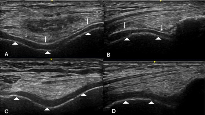
Ultrasonographic image in Double contour sign, suggestive of monosodium urate crystal deposition in hyaline cartilage.
Keywords:
Ultrasound, Double contour sign, GoutAbstract
Context: Microcrystalline arthropathies are diseases caused by deposition of crystals within joint and periarticular tissues. Three types of crystals involved in these diseases are monosodium urate, calcium pyrophosphate crystals, and basic calcium phosphate crystals (1). In the case of gout, hyaline cartilage is often the site of monosodium urate crystal deposits, revealing the "Double Contour" (DC) sign, which is a hyperechoic band over the chondrosynovial (superficial) margin, which increases the superficial cartilage interface to a thickness similar to that of subchondral bone (2). (Fig. 1).
Significance: The DC sign, along with other basic lesions such as tophi and aggregates, is a key ultrasound feature in the diagnosis of gout. The sensitivity and specificity of the DC sign vary between 42% and 87.8%, and 64.1% and 97%, respectively. The highest sensitivity is in the metatarsophalangeal joints, followed by the knees and metacarpophalangeal joints (3-4).
Combining the DC sign with power Doppler signals and elevated serum uric acid levels can increase the specificity for gout to 90% (3). Because of this, the double contour sign has been included in the new gout classification criteria proposed by the American College of Rheumatology and EULAR, reinforcing its importance in diagnosing this disease (5).
In conclusion, the double contour sign is a significant ultrasound indicator for diagnosing gout.
Figure 1: Right knee with double-contour sign (DC sign=white arrow; subchondral bone=arrowhead) in a longitudinal and transverse view of the hyaline cartilage (C and D) in a healthy knee, showing only hyaline cartilage and subchondral bone (arrowhead).
Publication Facts
Reviewer profiles N/A
Author statements
Indexed in
- Editor & editorial board
- profiles
- Academic society
- N/A

To learn about these publication facts, click ![]()
PF is maintained by the Public Knowledge Project
References
Filippucci E, Reginato AM, Thiele RG. Imaging of crystalline arthropathy in 2020. Best Pract Res Clin Rheumatol. 2020 Dec;34(6):101595.
Bruyn GA, Iagnocco A, Naredo E, Balint PV, Gutierrez M, Hammer HB et al. OMERACT Ultrasound Working Group. OMERACT Definitions for Ultrasonographic Pathologies and Elementary Lesions of Rheumatic Disorders 15 Years On. J Rheumatol. 2019 Oct;46(10):1388-1393.
Pârvănescu CD, Bărbulescu AL, Biță CE, et al. Ultrasound Features in Gout: An Overview. Med Sci (Basel). 2024;12(3):37.
Christiansen SN, Østergaard M, Slot O, Fana V, Terslev L. Ultrasound for the diagnosis of gout-the value of gout lesions as defined by the Outcome Measures in Rheumatology ultrasound group. Rheumatology (Oxford). 2021;60(1):239-249.
Richette P, Doherty M, Pascual E, Barskova V, Becce F, Castaneda J, et al. 2018 updated European League Against Rheumatism evidence-based recommendations for the diagnosis of gout. Ann Rheum Dis 2020;79:31–38.
Downloads
Published
How to Cite
Issue
Section
License
Copyright (c) 2025 Instituto Nacional de Rehabilitación Luis Guillermo Ibarra Ibarra

This work is licensed under a Creative Commons Attribution 4.0 International License.
© Instituto Nacional de Rehabilitación Luis Guillermo Ibarra Ibarra under a Creative Commons Attribution 4.0 International (CC BY 4.0) license which allows to reproduce and modify the content if appropiate recognition to the original source is given.